1/ Introduction and Historical Evolution
Microscopy began with the Nimrud lens (~700 BC), an Assyrian rock crystal used to magnify text and start fires. The first compound microscopes emerged in Europe around 1590, credited to Hans and Zacharias Janssen, with Galileo refining design shortly after and Anton van Leeuwenhoek later achieving up to 300X magnification with single lenses. Ernst Abbe’s 1873 work formalized resolution limits, anchoring modern optical theory.
2/ Fundamental Principles of Microscopy
Optical microscopy uses visible light focused by objective and eyepiece lenses to magnify specimens. Resolution is limited by diffraction; practical lateral resolution is on the order of 200nm under ideal conditions. Electron microscopy replaces photons with electron beams. In transmission electron microscopy (TEM), electrons pass through ultrathin sections to reveal sub-nanometer detail. In scanning electron microscopy (SEM), a focused electron beam rasters across the surface, generating topographical contrast.
3/ Classification and Key Modalities
Microscopes are broadly classified by illumination source and imaging mechanism:
- Optical (light) microscopes
- Electron microscopes (TEM, SEM)
- Scanning probe microscopes (AFM, STM)
- X-ray microscopes
Within optical microscopy, major modalities include:
- Bright-field: standard transmitted light
- Dark-field: scattered light imaging
- Phase-contrast: phase shifts convert to intensity contrast
- Differential interference contrast (DIC): enhances edges
- Fluorescence: specific wavelength excitation and emission
Super-resolution techniques (STED, SIM, PALM/STORM) overcome diffraction, achieving 20 - 50nm resolution.
4/ Core Components and Optical Design
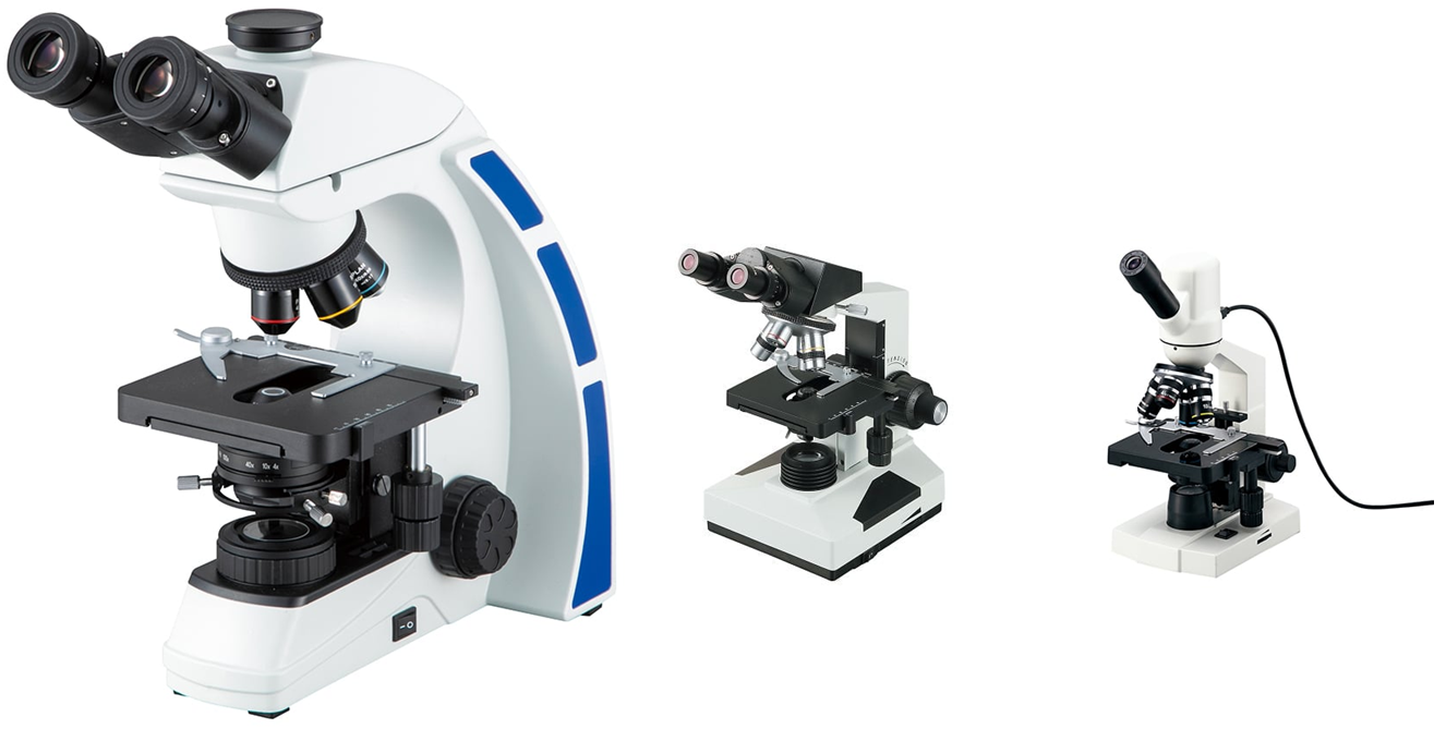
A compound light microscope comprises these primary subsystems:
- Illumination: Köhler alignment with halogen or LED source and condenser
- Objectives: multiple magnifications (4X - 100X) with defined numerical apertures
- Eyepieces: typically 10X or 15X magnification
- Mechanical stage: X-Y translation and precision focus knobs for coarse and fine adjustment
- Optical train: tube lens or relay lenses for infinity-corrected systems
- Camera port: integration with digital sensors or eyepiece adapters
5/ Sample Preparation Techniques
Achieving high-quality images depends on tailored sample prep:
- Fixation: crosslinking biomolecules with paraformaldehyde or glutaraldehyde
- Embedding and sectioning: paraffin for histology; epoxy for TEM; cryo-microtomy for native states
- Staining: H&E, silver stains for light microscopy; immunogold or heavy-metal stains for TEM; fluorescent dyes or antibodies for fluorescence
- Cryo-preservation: vitrification to avoid ice crystals in cryo-EM
6/ Advanced Imaging and Image Processing
Modern workflows integrate hardware and software:
- Confocal and multiphoton microscopy enable optical sectioning and deep-tissue imaging
- Deconvolution algorithms reverse diffraction blur, improving contrast
- Image stitching creates large-field mosaics from tiled captures
- Quantitative analysis: automated segmentation and intensity measurements via open-source tools (e.g., ImageJ, CellProfiler)
- AI-assisted super-resolution uses deep learning to predict sub-diffraction details
7/ Applications Across Disciplines
Microscopy underpins breakthroughs in multiple fields:
- Life sciences: cellular dynamics, pathogen identification, developmental biology
- Materials science: nanoparticle characterization, thin-film analysis, crystal defects
- Semiconductor industry: defect inspection, metrology of microelectronic components
- Environmental monitoring: nanopollutant detection, microplastic analysis
8/ Maintenance, Calibration, and Quality Control
Regular upkeep ensures optimal performance:
- Clean optics with lens tissue and solvent; avoid abrasive contact
- Align illumination and condenser annually or after major movement
- Calibrate magnification and stage micrometer for accurate measurements
- Service light sources and stage mechanics per manufacturer schedules
9/ Emerging Trends and Future Directions
The frontiers of microscopy include:
- Correlative Light and Electron Microscopy (CLEM) for multiscale imaging
- Cryo-EM single-particle analysis delivering near-atomic models of macromolecules
- Quantum microscopy exploiting photon entanglement to break traditional limits
- Portable, open-source microscopes using 3D-printed optics and smartphone integration
- AI-driven autonomous platforms for high-throughput screening and diagnostics
Visit qtetech.com/en to choose the right microscope for you from famous brands.
QTE Technologies is proud to be a global MRO provider, serving customers in over 180 countries and always strives to ensure customers have a complete and satisfied experience. We were established in 2010, providing over 1 million products for every industry and science and technology. In addition, you can contact us anytime via 24×7 chat support, phone, WhatsApp or email. Discover what our valued customers say about our services on our dedicated review page.
Author: Editorial Board of QTE Technologies (with a strong background in both engineering and innovation - accumulated over 15 years of experience).